DARA Institute is a dynamic organization dedicated to strengthening individuals and communities through practical education, skills development, and research-driven solutions. By integrating multiple sectors such as technology, health, governance, and livelihoods, it delivers hands-on training and evidence-based programs that promote sustainable development. Working in partnership with governments, NGOs, and private sector actors, the Institute focuses on creating inclusive, real-world impact and long-term social and economic growth.
DARA Institute is a dynamic organization dedicated to strengthening individuals and communities through practical education, skills development, and research-driven solutions. By integrating multiple sectors such as technology, health, governance, and livelihoods, it delivers hands-on training and evidence-based programs that promote sustainable development. Working in partnership with governments, NGOs, and private sector actors, the Institute focuses on creating inclusive, real-world impact and long-term social and economic growth.
Diagnostic ultrasonography is the most operator-dependent imaging discipline in medicine. Two clinicians scanning the same patient on the same machine can reach entirely different conclusions, because the diagnosis is created at the moment of scanning — not afterwards, in front of a screen. Competence therefore requires far more than knowledge of pathology.
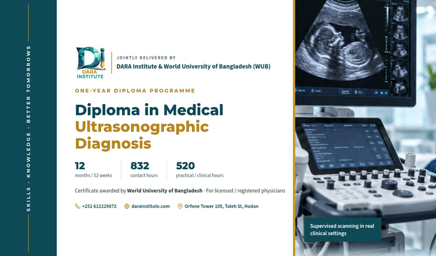
The Diploma in Medical Ultrasonographic Diagnosis is a full one-year, competency-based programme that trains licensed physicians to perform and interpret general diagnostic ultrasound examinations independently, safely and within a clearly defined scope of practice. It is built around a simple progression:
Knowledge → Technical skill → Image interpretation → Clinical reasoning → Reporting → Professional practice
Over 52 weeks, trainees move from ultrasound physics and machine handling to complete abdominal, genitourinary, pelvic, obstetric, small-parts, vascular, emergency, thoracic, musculoskeletal and paediatric examinations — supported throughout by supervised scanning on real patients, weekly case conferences, a mandatory logbook and a supervised scholarly project.
The programme is deliberately designed around competency rather than attendance. Sitting through the sessions is not sufficient to graduate: every trainee must accumulate documented, signed-off examinations at an independent level of performance before the final award is made.
Programme Aim
To produce physicians who can independently perform and interpret appropriately selected general diagnostic ultrasound examinations within their demonstrated scope of competence, while recognising diagnostic limitations and referring examinations that require specialist expertise.
Who Should Apply?
General practitioners and family physicians who need bedside diagnostic imaging in daily practice
Emergency and critical-care physicians seeking structured FAST/eFAST and point-of-care competence
Obstetricians, gynaecologists and reproductive health clinicians
Internal medicine and surgical clinicians managing abdominal and hepatobiliary disease
Paediatricians and neonatal clinicians
Hospital and health-centre medical officers working where no radiologist is on site
Physicians in NGO, humanitarian and ministry health facilities who carry diagnostic responsibility alone
Doctors preparing for further specialisation in radiology or imaging-intensive disciplines
Why Choose This Diploma?
Competency-based, not attendance-based. Graduation depends on documented, signed-off performance — not on hours sitting in a classroom.
Approximately 520 hours of supervised practical and clinical training — about 62.5% of all contact time is spent with a probe in hand.
A target of around 900 documented scan encounters, deliberately set well above the minimum needed simply to pass, so that trainees see normal anatomy, common disease and technically difficult patients repeatedly.
Small-group hands-on teaching at a preferred ratio of one instructor to four–six trainees, with a maximum cohort of 20.
Real patients in real clinical settings, with rotations in general, obstetric and gynaecologic, vascular and advanced ultrasound.
International benchmarking against AIUM, EFSUMB, ISUOG and WFUMB educational standards.
Complete diagnostic training, not just image acquisition — clinical reasoning, differential diagnosis, structured reporting and referral decisions are examined in their own right.
Weekly clinical case conferences with technical critique, pathology correlation and reporting review.
A supervised scholarly capstone — an audit, diagnostic study or quality-improvement project rooted in the trainee’s own practice.
Dual institutional strength: delivered by DARA Institute with academic supervision and certification from the World University of Bangladesh.
Entry Requirements
MBBS or an equivalent primary medical qualification.
Current registration with the relevant national medical regulatory authority, or equivalent registration for international applicants where legally applicable.
Ability to communicate effectively in English.
Basic computer literacy.
Successful completion of the DARA Institute admission screening and interview, where applicable.
Note: no prior ultrasound experience is required. The programme begins with physics, instrumentation and machine handling and builds from first principles. Final eligibility requirements are aligned with the recognition and accreditation authority under which the programme is registered.
Learning Outcomes
By the end of the programme, graduates will be able to:
Knowledge
Explain ultrasound physics and instrumentation
Explain Doppler principles
Describe ultrasound bioeffects and safety, and apply the ALARA principle
Describe normal sonographic anatomy
Explain the relevant pathophysiology
Recognise the common sonographic manifestations of disease
Technical skills
Select an appropriate transducer and position the patient correctly
Optimise machine settings
Perform standardised examinations and acquire complete image sets
Perform basic and advanced Doppler studies appropriate to the examination
Recognise technical artefacts and troubleshoot poor-quality examinations
Diagnostic competence
Identify normal anatomy and detect abnormal findings
Characterise lesions and form appropriate differential diagnoses
Integrate ultrasound findings with clinical information
Recognise urgent findings
Identify situations in which ultrasound is insufficient and further imaging or referral is required
Professional competence
Produce structured diagnostic reports and communicate significant findings appropriately
Maintain a clinical logbook and participate in quality-assurance activities
Maintain patient confidentiality and demonstrate professional and ethical conduct
Understand scope-of-practice limitations
Develop habits of lifelong learning and continuing professional competence
$50 / Month
This Course Included
Term
Oct 2026 - Oct 2027
Dates
01 Oct 2026 –
01 Oct 2027
Duration
12 Months / 52 Weeks
Duration: 12 months / 52 weeks
Academic structure: Four parts × three months
Contact time: Approximately 832 hours (16 hours per week)
Practical / clinical education: Approximately 520 hours
Clinical case exposure: Approximately 900 documented scan encounters
Training mode: Face-to-face, supervised clinical and practical training
Maximum intake: 20 trainees per cohort
Logbook and capstone: Both mandatory
Final examination: Written + image interpretation + OSCE + clinical/practical + viva voce
Certification: World University of Bangladesh, Centre for Professional Development
Language
English
Skill Level
Licensed Medical Doctors / Postgraduate Professional
The programme consists of four sequential parts, each assessed before progression.
Part
Months
Principal theme
Weeks
Scan target
Part I
1–3
Foundations of Diagnostic Ultrasonography
1–13
201
Part II
4–6
Comprehensive General Ultrasonography
14–26
240
Part III
7–9
Obstetric, Gynaecologic and Doppler Ultrasonography
27–39
265
Part IV
10–12
Advanced Ultrasound and Clinical Integration
40–52
215
Part I — Foundations of Diagnostic Ultrasonography (Weeks 1–13)
Physics, instrumentation, safety, machine handling and normal abdominal anatomy. Trainees learn to produce a technically adequate image before they are asked to interpret one.
Week
Lecture / seminar
Practical and clinical session
Learning outcome
Scan target
1
Introduction to ultrasound: indications, limitations, terminology
Part I scan target: approximately 201 examinations / scan encounters.
Part II — Comprehensive General Ultrasonography (Weeks 14–26)
Systematic coverage of abdominal, hepatobiliary, renal and genitourinary, gastrointestinal, pelvic, breast, small-parts and emergency ultrasound, with pathology correlation throughout.
Week
Lecture / seminar
Practical and clinical session
Learning outcome
Scan target
14
Liver pathology I
Fatty liver, hepatitis, cirrhosis
Recognise diffuse liver disease
20
15
Liver pathology II
Focal liver lesions
Characterise common lesions
20
16
Gallbladder and biliary disease
Stones, sludge, cholecystitis
Diagnose common biliary pathology
20
17
Pancreatic disease
Pancreatitis, masses, cysts
Recognise pancreatic pathology
15
18
Spleen and portal system
Splenomegaly and portal Doppler
Evaluate portal hypertension
15
19
Renal pathology
Stones, cysts, masses, obstruction
Diagnose common renal abnormalities
20
20
Bladder and prostate
Bladder, post-void residual, prostate
Perform a genitourinary examination
20
21
Gastrointestinal ultrasound
Bowel, appendix, obstruction
Recognise bowel pathology
15
22
Pelvic ultrasound
Transabdominal pelvis
Evaluate the uterus and adnexa
20
23
Gynaecologic pathology
Fibroids, adenomyosis, ovarian lesions
Characterise common pathology
20
24
Breast ultrasound
Normal breast and common lesions
Perform a systematic breast examination
15
25
Thyroid, neck and scrotum
Small-parts scanning
Evaluate common superficial pathology
20
26
Emergency ultrasound
FAST/eFAST, AAA, acute abdomen
Perform an emergency examination
20
Part II scan target: approximately 240 examinations / scan encounters.
Part III — Obstetric, Gynaecologic and Doppler Ultrasonography (Weeks 27–39)
First, second and third trimester obstetric scanning, basic fetal anomaly screening, obstetric Doppler, advanced gynaecology and vascular Doppler, benchmarked against ISUOG practice guidance.
Week
Lecture / seminar
Practical and clinical session
Learning outcome
Scan target
27
Early pregnancy
Gestational sac, yolk sac, embryo
Assess early pregnancy
20
28
Pregnancy failure and ectopic pregnancy
Early pregnancy cases
Recognise ectopic and non-viable pregnancy patterns
20
29
First-trimester dating
Crown-rump length and viability
Perform accurate dating
20
30
11–14 week assessment
Nuchal translucency and early anatomy
Perform basic first-trimester screening
20
31
Normal second trimester
Fetal biometry
Obtain standard fetal measurements
20
32
Normal third trimester
Growth and fluid assessment
Assess fetal growth
20
33
Placenta, cervix and amniotic fluid
Placental and cervical assessment
Identify praevia and fluid abnormalities
20
34
Fetal CNS and face
Neurosonography basics
Recognise major CNS and facial abnormalities
20
35
Fetal thorax and abdomen
Anomaly scanning
Recognise major structural anomalies
20
36
Fetal heart
Four-chamber, outflow tracts, three-vessel view
Perform basic cardiac screening
20
37
Obstetric Doppler
Umbilical, middle cerebral and uterine artery
Perform and interpret basic Doppler
20
38
Advanced gynaecology
Endometrium, adnexa, endometriosis, infertility
Integrate pelvic findings
20
39
Vascular Doppler
Carotid, DVT, arterial and venous studies
Perform basic vascular ultrasound
25
Part III scan target: approximately 265 examinations / scan encounters.
Part IV — Advanced Ultrasound and Clinical Integration (Weeks 40–52)
Advanced Doppler, thyroid and neck, thoracic, musculoskeletal, paediatric and point-of-care ultrasound, together with structured reporting, differential diagnosis, quality assurance and a final integrated clinical rotation.
Week
Lecture / seminar
Practical and clinical session
Learning outcome
Scan target
40
Advanced abdominal Doppler
Hepatic, portal and renal Doppler
Interpret vascular physiology
20
41
Advanced thyroid and neck
Nodules and lymph-node mapping
Risk-stratify lesions
15
42
Thoracic ultrasound
Lung, pleura, diaphragm
Recognise common thoracic pathology
15
43
Musculoskeletal ultrasound
Shoulder, knee, soft tissue
Perform basic MSK examinations
15
44
Paediatric and neonatal ultrasound
Paediatric abdomen and neurosonography
Recognise common paediatric findings
15
45
POCUS and emergency integration
Shock, dyspnoea, trauma
Apply focused ultrasound appropriately
20
46
Ultrasound-guided procedures
Needle visualisation and simulation
Demonstrate safe needle visualisation
10
47
Structured reporting
Reporting laboratory
Produce complete diagnostic reports
20
48
Differential diagnosis
Unknown-case laboratory
Build evidence-based differentials
20
49
Quality assurance
Audit and peer review
Perform QA and identify errors
15
50
Integrated clinical rotation
Independent supervised scanning
Manage complete examinations
25
51
Final clinical consolidation
Final logbook cases
Demonstrate independent competence under supervision
25
52
Final examinations
OSCE, viva, image interpretation
Demonstrate graduation competence
Part IV scan target: approximately 215 examinations / scan encounters.
Clinical Training and Case Volume
The curriculum targets approximately 900 documented examinations across the year. The number is deliberately higher than the minimum exposure required simply to pass a course: it is set to give repeated exposure to normal anatomy, to common disease and to technically difficult examinations.
How examinations are classified
Category
Definition
Counts toward final independent portfolio
A — Observed
The trainee observes the instructor.
No
B — Assisted
The trainee performs portions of the examination with direct instructor assistance.
No
C — Supervised
The trainee performs the examination while the instructor is available.
Yes
D — Competency-certified
The trainee independently performs the examination and the supervisor signs competency.
Yes
Only categories C and D count toward the final independent competency portfolio.
Minimum competency case distribution
Domain
Minimum documented cases
Minimum competency sign-offs
Complete abdomen
150
30
Hepatobiliary
100
20
Renal / genitourinary
100
20
Gastrointestinal
40
10
Pelvis / gynaecology
60
15
Obstetrics
150
30
First trimester
40
10
Second / third trimester
80
20
Obstetric Doppler
30
10
Vascular Doppler
75
15
Thyroid / neck
40
10
Breast
30
8
Scrotum / small parts
25
6
Emergency / POCUS
40
10
Thoracic
20
5
Musculoskeletal
20
5
Paediatric / neonatal
20
5
Other / special cases
30
Approximate total
1,030
—
These are DARA Institute programme targets rather than assertions that any single international organisation mandates this exact number. Final case requirements are reconciled with the requirements of the recognition body and with specialty-specific standards, and remain subject to clinical case availability and specialty distribution.
Competency levels
C1 — Knowledge: Can describe the indication, anatomy, technique, pathology and limitations.
C2 — Assisted performance: Can perform the examination with significant instructor assistance.
C3 — Supervised performance: Can complete the examination under supervision.
C4 — Independent competency: Can independently select the protocol, obtain complete images, optimise the examination, perform appropriate Doppler, recognise pathology, formulate an impression and report the findings.
C5 — Advanced competency: Can manage technically difficult cases and recognise when additional imaging, consultation or referral is required.
Graduation requires C4 competency in the core examination domains.
Teaching is face-to-face and practice-led. The programme uses lectures, interactive seminars, image-based teaching, live demonstrations, supervised scanning, real-patient training, case-based and problem-based learning, pathology correlation, reporting workshops, simulation where available, OSCE practice, peer review, clinical audit and independent study.
Recommended weekly pattern
Session
Focus
Hours
Session 1
Theory
2
Session 2
Interactive / image teaching
2
Session 3
Demonstration
2
Session 4
Hands-on scanning
4
Session 5
Clinical scanning
4
Session 6
Case and report conference
2
Total
Contact hours per week
16
Across 52 weeks this gives approximately 832 contact hours — about 312 theoretical and 520 practical or clinical. Independent reading, case preparation, logbook completion, research and capstone work occur in addition to scheduled contact hours.
Faculty-to-trainee ratios
Activity
Ratio
Didactic teaching
Maximum 1:20
Image interpretation
Maximum 1:10
Hands-on scanning
Preferred 1:4–6
OSCE
Approximately 1 examiner : 1 candidate per station
Weekly clinical case conference
At least one case conference is held every week. Each session covers the clinical presentation, the ultrasound images, a technical critique, the differential diagnosis, the final diagnosis, pathology or radiology correlation where available, the management implication, and a critique of the report.
Supervision and feedback
Monthly: a clinical competency report, a case-volume report, documented strengths, documented deficiencies and a remediation plan where needed.
Quarterly: a formal progress review.
Final: a comprehensive competency report.
Assessment Framework
Assessment is continuous and multimodal. No single examination determines the outcome.
Assessment component
Weight
Part I examination
10%
Part II examination
10%
Part III examination
10%
Continuous clinical competency
15%
Logbook
10%
Image interpretation
10%
OSCE
15%
Final theory examination
10%
Final clinical / practical examination
5%
Viva voce
2.5%
Dissertation / capstone
2.5%
Total
100%
Formative assessment
Formative assessment runs continuously through weekly image quizzes, supervised scanning checklists, monthly case reviews, report-writing exercises, Doppler interpretation exercises and clinical case presentations. Formative assessments may identify remediation needs but do not necessarily contribute directly to final marks.
Final theory examination
Paper 1 — Ultrasound Science (100 marks): physics, instrumentation, Doppler, artefacts, safety and quality assurance.
Paper 2 — Clinical Ultrasound (100 marks): abdomen, gastrointestinal, genitourinary, pelvis, breast, thyroid, scrotum, emergency, obstetrics and gynaecology, vascular, thoracic and musculoskeletal ultrasound.
Written examination blueprint
Domain
Weight
Physics and instrumentation
15%
Doppler
10%
Safety and quality assurance
5%
Abdomen, gastrointestinal and genitourinary
25%
Pelvis and gynaecology
10%
Obstetrics
20%
Small parts, breast and thyroid
7.5%
Emergency, thoracic, musculoskeletal and POCUS
7.5%
Total
100%
Questions are distributed across cognitive levels — approximately 20% recall, 25% understanding, 30% application and 25% analysis and clinical reasoning. At least 55% of the clinical paper requires application or analysis rather than simple recall.
Image interpretation examination
Fifty cases, each containing a clinical history, representative images, measurements where appropriate and Doppler images where applicable. For each case the candidate must give the key finding, the interpretation, a differential, the most likely diagnosis and a recommendation where appropriate.
Cases span normal anatomy, common pathology, uncommon but important pathology, artefacts, technically inadequate examinations, Doppler, obstetric and emergency studies. At least 20% of cases test recognition of limitations or of technically inadequate studies.
OSCE
Twelve stations of eight minutes each:
Station
Content
1
Machine operation and optimisation
2
Probe selection and image orientation
3
Abdominal examination
4
Hepatobiliary pathology
5
Renal and genitourinary examination
6
Thyroid and small-parts examination
7
Obstetric biometry
8
First-trimester ultrasound
9
Obstetric Doppler
10
Vascular / DVT examination
11
Image interpretation and differential diagnosis
12
Report writing and clinical communication
Each station carries 20 marks, for a total of 240. Marks within a station are distributed across patient safety (2), preparation (2), technique (6), image quality (3), interpretation (4) and communication or documentation (3).
Final clinical examination
The candidate receives a real patient and must review the clinical indication, select the examination, explain it to the patient, perform the scan, optimise the images, obtain a complete image set, perform relevant Doppler, identify the abnormality, provide a differential, formulate an impression and write the report.
Viva voce
The viva covers physics, artefacts, pathology, differential diagnosis, Doppler, safety, limitations, reporting, clinical correlation and referral decisions. It emphasises clinical reasoning, not rote memorisation.
Dissertation / Capstone Project
Each trainee completes one supervised scholarly project of 5,000–8,000 words, structured as: title, background, research question, objectives, literature review, methodology, ethical considerations, results, discussion, limitations, conclusion, recommendations and references.
Accepted designs, with worked examples:
Clinical audit — completeness of routine abdominal ultrasound reports before and after implementation of a structured reporting template.
Diagnostic study — diagnostic performance of ultrasound for suspected acute cholecystitis.
Retrospective study — sonographic characteristics of thyroid nodules in a tertiary-care population.
Prospective study — ultrasonographic findings in patients presenting with suspected renal colic.
Quality improvement — reduction of technically incomplete ultrasound examinations following introduction of a competency-based scanning checklist.
Progression, Pass Marks and Professional Standards
Pass / fail criteria
A candidate passes the programme only when both the academic and the clinical requirements are fulfilled:
Overall final score ≥ 50%
Final theory examination ≥ 50%
Image interpretation examination ≥ 50%
OSCE ≥ 60%
Final clinical / practical examination ≥ 60%
Viva ≥ 50%
Capstone ≥ 50%
Required clinical logbook completed
Required core competency sign-offs completed
No unresolved patient-safety competency failure
Critical-competency rule
A candidate may not graduate merely by achieving an aggregate pass mark if they demonstrate serious incompetence in a safety-critical domain. Automatic remediation is required for serious failure involving incorrect patient identification, unsafe examination, failure to recognise major emergency pathology, grossly inappropriate Doppler technique, serious breach of infection control, falsification of records, inability to obtain basic required examinations, or serious professional misconduct.
Remediation
A candidate failing a competency receives documented feedback, additional supervised training, targeted case allocation and repeat assessment. The maximum recommended remediation period is 8–12 weeks. A candidate may be required to repeat a clinical rotation if competency remains inadequate.
Attendance
A minimum of 90% of scheduled theoretical sessions and 95% of mandatory clinical and practical sessions is required. Absence from a critical practical competency session must be made up. Attendance alone does not constitute successful completion.
Professional conduct
Patient confidentiality, informed consent, respectful communication, appropriate professional attire, infection-control compliance, accurate documentation, truthful logbook entries and academic integrity are mandatory throughout. Fabrication of clinical cases, images or logbook entries constitutes serious academic misconduct.
Logbook and Competency Portfolio
Completed logbook and core competency forms
Minimum case volume achieved
20 representative complete examinations
20 pathology cases
10 Doppler cases
10 obstetric cases
5 emergency cases
5 difficult or technically challenging cases
Five supervised reports and five QA / audit cases
Capstone / dissertation
Faculty evaluations and the final competency declaration
Minimum representative image portfolio
Domain
Cases
Domain
Cases
Abdomen
5
Doppler
3
Genitourinary
3
Thyroid / small parts
2
Pelvis
3
Emergency
2
Obstetrics
5
Total
23 complete studies
Certification and Graduate Profile
Certificates are issued for each of the four parts, leading to the full diploma award. The certificate does not simply state that the holder completed a course in ultrasound; subject to the approved wording of the recognition authority, it states:
“The holder has successfully completed the competency-based Certificate in Medical Ultrasonographic Diagnosis (CMUD), including structured theoretical education, supervised clinical scanning, diagnostic image interpretation, Doppler ultrasonography, obstetric and gynecologic ultrasonography, structured reporting, quality assurance and competency-based clinical assessment.”
Scope of practice: the certificate does not imply specialist status or authorisation beyond the graduate’s medical licence, training and institutional scope of practice.
Graduate profile
The successful graduate functions as a competent physician-level general diagnostic ultrasonography practitioner within the scope defined by the programme and by applicable professional regulations, with particular competence in:
Abdominal, hepatobiliary and renal / genitourinary ultrasound
Pelvic ultrasound and basic transvaginal ultrasound
Obstetric ultrasound, basic fetal anomaly screening and obstetric Doppler
Gynaecologic ultrasound
Thyroid and neck, breast and scrotal ultrasound
Basic vascular Doppler
Emergency ultrasound and basic thoracic ultrasound
Introductory musculoskeletal ultrasound
Structured reporting and quality assurance
The graduate is also able to recognise examinations that require specialist referral or additional imaging.
Recommended core textbooks
Area
Reference
General ultrasound
Rumack CM et al. Diagnostic Ultrasound
General ultrasound
Middleton WD et al. Ultrasound: The Requisites
General ultrasound
Hagen-Ansert SL. Textbook of Diagnostic Sonography
Physics
Kremkau FW. Diagnostic Ultrasound: Principles and Instruments
Obstetrics and gynaecology
Callen PW. Ultrasonography in Obstetrics and Gynecology
Obstetrics and gynaecology
ISUOG educational resources and practice guidelines
Vascular
Zwiebel WJ. Introduction to Vascular Ultrasonography
Musculoskeletal
Bianchi S, Martinoli C. Ultrasound of the Musculoskeletal System
Intake, Fees and How to Apply
Course Term
Oct 2026 - Oct 2027
Start Date
1 October 2026
Duration
12 months / 52 weeks
Cohort Size
Maximum 20 trainees
Delivery
Face-to-face with supervised clinical and practical training
Venue
DARA Institute training centre, Orfone Tower 105, Taleh Street, Hodan, Mogadishu, with affiliated clinical training sites
Tuition
To be confirmed by DARA Institute admissions
Application steps
Complete the online registration form on the DARA Institute website, or visit the training centre.
Submit your medical qualification, current registration certificate and a copy of your identification.
Attend the admission screening and interview.
Confirm your place and complete enrolment before the cohort start date.
Frequently Asked Questions
Do I need previous ultrasound experience?
No. The programme starts with ultrasound physics, instrumentation and machine handling and builds from first principles. What you do need is a primary medical qualification and current registration.
Can I take the programme online or part-time?
No. Supervised scanning on real patients is the core of the diploma, and roughly 62.5% of contact time is practical or clinical. The clinical component is mandatory and cannot be delivered remotely.
How many scans will I actually perform?
The programme targets approximately 900 documented scan encounters across the year, subject to clinical case availability. Only examinations performed under supervision or independently with a supervisor sign-off count toward the final competency portfolio.
What happens if I fail a component?
You receive documented feedback, additional supervised training, targeted case allocation and a repeat assessment, normally within 8–12 weeks. If competency remains inadequate, a clinical rotation may have to be repeated.
Is attendance enough to graduate?
No. Attendance minimums exist (90% theory, 95% mandatory clinical sessions), but the award depends on documented competency sign-offs, a completed logbook and performance in the final examinations.
Who awards the certificate?
The certificate is awarded by the World University of Bangladesh through its Centre for Professional Development, with the programme delivered by DARA Institute.
Does the diploma make me a radiologist or a specialist?
No. It certifies competency-based training in general diagnostic ultrasonography. It does not imply specialist status or authorisation beyond your medical licence, your training and your institutional scope of practice.
What is the capstone project?
A supervised scholarly project of 5,000–8,000 words — an audit, a diagnostic study, a retrospective or prospective study, or a quality-improvement project — usually drawn from your own clinical practice.